Vadzo Imaging has positioned the Innova-234CGE, a 2MP global shutter GigE camera built on the Onsemi AR0234 sensor, for digital pathology and clinical laboratory imaging systems. The camera pairs global shutter capture with a native GigE Vision interface, addressing the motion artifact, cable length, and stage synchronization requirements that whole slide scanning and microscopy platforms depend on. This positioning reflects growing demand from pathology instrument builders for a camera that holds a moving slide image steady, connects over standard Ethernet cabling across a lab bench, and synchronizes precisely with a motorized microscope stage.
FORT WORTH, TX / ACCESS Newswire / September 29, 2026 / Vadzo Imaging, a provider of embedded vision camera products, today announced the positioning of the Innova-234CGE AR0234 Global Shutter GigE Camera for digital pathology and clinical laboratory imaging systems. The Innova-234CGE is built on the Onsemi AR0234 sensor and joins Vadzo’s GigE Vision camera portfolio. It combines global shutter capture with a native Gigabit Ethernet interface in a single camera module. This positioning targets pathology instrumentation where a camera must hold a slide image free of motion distortion while a motorized stage scans across the specimen and must connect to a host computer over a longer cable run than USB typically allows. With this positioning, Vadzo delivers an AR0234 2MP Gigabit Ethernet Camera built specifically for the imaging demands of digital pathology and life sciences laboratory instruments.
The Digital Pathology Imaging Problem
Digital pathology and whole slide scanning systems place a camera in a role that few consumer or general-purpose industrial sensors are built for. The camera sits behind a microscope objective, captures a moving specimen as a motorized stage scans across the slide, and must deliver diagnostic-quality color at every tile the scanner stitches into a final image. Instrument manufacturers evaluating a sensor for this role quickly find that a camera built for a static industrial inspection line does not automatically translate to a moving stage scanning environment, where timing, motion, and color all have to hold up across thousands of captures per slide. Three problems come up again and again when instrument builders select a camera for this application.
Motion distortion during stage scanning is the first. Whole slide scanners move the specimen stage continuously or in rapid steps to capture the full tissue area at high resolution, and a rolling shutter sensor exposes each row of pixels at a slightly different moment during that motion. A Global Shutter Pathology Camera avoids this entirely because the sensor captures every pixel at the same instant, which keeps cell boundaries, nuclei, and tissue structures sharp even while the stage is actively moving between fields of view.
Cable length and electrical noise are the second problem. Many pathology and laboratory imaging setups place the camera on a microscope stand while the workstation sits at a separate bench position, sometimes several meters away to keep vibration and electrical interference away from the optical path. A USB camera is typically limited to a few meters of unpowered cable run before signal integrity degrades. A GigE Pathology Vision Sensor connects over standard Ethernet cabling that reliably reaches up to 100 meters, giving lab designers far more flexibility in bench layout than a USB interface allows.
Stage and trigger synchronization is the third problem. A slide scanner has to fire the camera at the exact moment the stage reaches each imaging position, and any timing drift between stage motion and camera exposure produces blurred or misaligned tiles that break the stitching algorithm used to assemble the final whole slide image. A Global Shutter GigE Lab Sensor with hardware trigger input lets the stage controller fire the camera at a precise, repeatable moment rather than relying on software polling that introduces timing uncertainty.
A Pathology GigE Sensor Kit that solves all three problems inside one validated camera module removes a significant engineering burden from instrument builders who would otherwise need to qualify a sensor, a cabling strategy, and a synchronization method separately before a scanner design is ready for production. That separation of concerns, treating imaging, connectivity, and timing as three independent qualification tasks, is exactly what a single validated camera platform is designed to eliminate.
Why Global Shutter and GigE Vision Belong Together for Pathology
The AR0234 Global Shutter GigE Camera pairs two engineering elements that matter specifically to laboratory imaging instrumentation. Vadzo selected the Onsemi AR0234 sensor for its global shutter architecture and paired it with a GigE Vision interface because pathology and microscopy platforms need both distortion-free capture and industrial-grade connectivity, a combination that consumer imaging sensors and USB interfaces were never designed to deliver together. Many camera vendors offer one half of this pairing, a global shutter sensor without a laboratory grade interface, or a GigE Vision camera built around a rolling shutter sensor that was never intended for a moving stage application.
Global shutter capture is the imaging foundation. Every pixel on the Onsemi AR0234 sensor exposes at the same instant, which is what allows a Global Shutter Ethernet Slide Sensor to hold a clean image of tissue morphology even as the stage moves the specimen through the field of view during a scan. This matters directly to diagnostic image quality, since distorted or smeared cell boundaries can obscure the fine structural detail a pathologist relies on when reviewing a digitized slide.
GigE Vision connectivity is layered on top of that imaging foundation. The AR0234 Global Shutter GigE Camera streams standardized GigE Vision data over Gigabit Ethernet, which brings long cable reach, optional Power over Ethernet, and a widely supported GenICam control interface that laboratory software vendors already know how to integrate against. A Global Shutter Lab Vision Sensor built on an open standard like GigE Vision avoids locking an instrument builder into a proprietary driver stack that could complicate long-term software support.
Trigger and timing control close the loop. A 2MP Global Shutter Pathology Sensor with GenICam-exposed hardware trigger inputs lets a scanner’s motion controller fire image capture at a precisely repeatable moment relative to stage position, which is the foundation that tile stitching algorithms depend on to assemble accurate, artifact-free whole slide images from hundreds or thousands of individual captures.
Product Overview
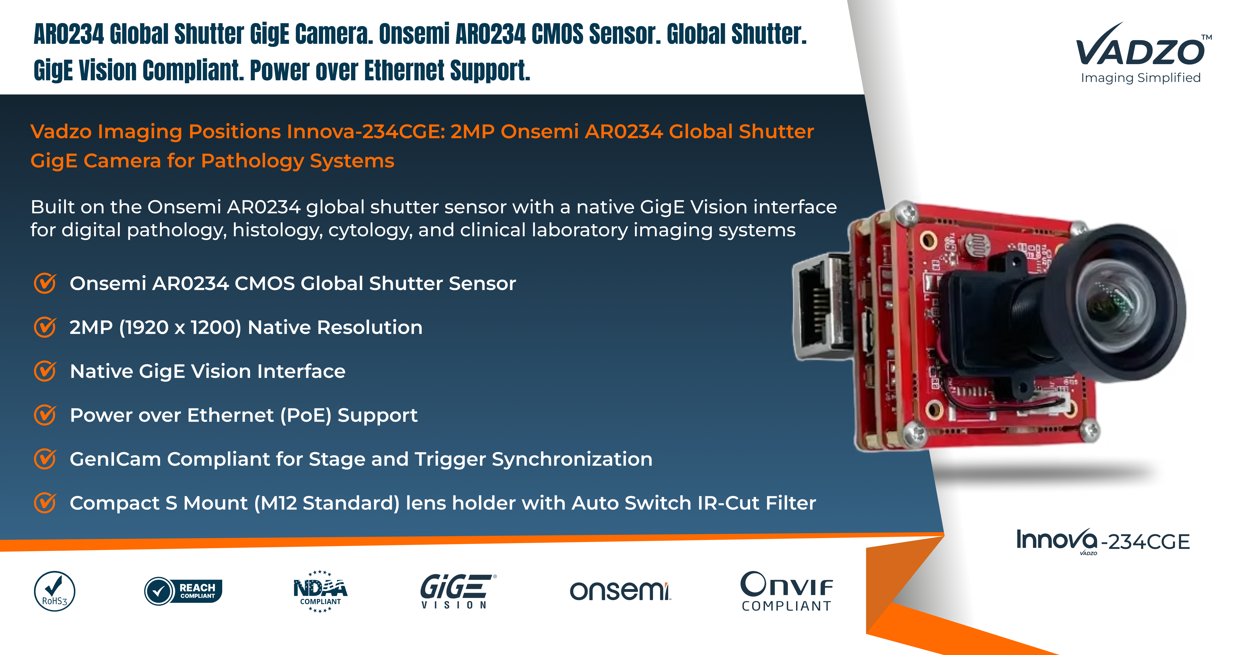
The Innova-234CGE is a 2MP Pathology Ethernet Camera built on the Onsemi AR0234 CMOS sensor and engineered for digital pathology and laboratory imaging instrumentation. The AR0234 is a 2MP (1920 x 1200) global shutter sensor with a 1/2.6 inch optical format and a 3.0-micron pixel that delivers accurate color reproduction, a requirement for interpreting the staining patterns pathologists rely on when reviewing H&E and other stained tissue sections. Frame rates up to 120 fps at reduced resolution support the throughput that high-speed scanning workflows need.
The Innova-234CGE connects over a native GigE Vision interface with GenICam-compliant control, presenting a standard camera interface to any imaging software written against the GigE Vision standard. As an AR0234 Global Shutter Gigabit Ethernet Camera, the module supports Power over Ethernet, letting a single Ethernet cable carry both data and power to simplify cabling in a crowded laboratory bench environment. The camera ships with an S-Mount optical interface, the standard mount for microscope camera ports, so it integrates directly onto existing trinocular microscope heads and slide scanning optical trains without a custom adapter.
Vadzo Imaging positioned the Innova-234CGE for pathology systems after engaging with instrument builders who described the same recurring qualification challenge: sourcing a camera with proven global shutter performance, validated GigE Vision compliance, and a color response tuned for stained tissue imaging, all from a single vendor rather than assembling a solution from mismatched components. The result is a camera module that laboratory instrument teams can specify with confidence that the sensor, interface, and optical mount all work together as a coherent system. This matters particularly for medical device manufacturers who need to document their camera selection rationale as part of a broader instrument design history file, where a camera with clearly published specifications and a stable long-term supply relationship reduces downstream risk compared to sourcing components from multiple vendors with inconsistent documentation.
Key specs: 2MP (1920 x 1200) | Onsemi AR0234 CMOS, 1/2.6-inch, 3.0-micron pixel | Color | Global Shutter | GigE Vision with PoE | GenICam Compliant | S Mount (M12 Standard) with Auto Switch IR-Cut Filter
Key Capabilities of the Innova-234CGE Global Shutter GigE Camera
Global Shutter Imaging for Moving Slide Capture
Rolling shutter sensors expose a frame row by row, which distorts any subject that moves during the exposure window. On a whole slide scanner where the stage is continuously repositioning the specimen, that distortion appears as skewed cell boundaries and blurred tissue edges. The Onsemi AR0234 sensor inside the Innova-234CGE exposes the entire frame at once, so a 2MP Ethernet Lab Camera built on this sensor holds tissue morphology sharp even during active stage motion. A 2MP Pathology Sensor Module of this type is what allows automated tile stitching software to assemble a clean whole slide image without visible seams caused by frame-to-frame distortion. This distinction becomes more pronounced as scanning speed increases, since faster stage movement gives a rolling shutter sensor even less time to complete a clean row-by-row exposure before the next field of view arrives.
GigE Vision Interface for Long Cable Runs
An Onsemi AR0234 Pathology Camera frequently sits on a microscope stand positioned away from the workstation to isolate the optical path from vibration and electrical noise. The Innova-234CGE connects over standard Gigabit Ethernet cabling that reliably supports runs up to 100 meters, far beyond what an unpowered USB connection can reliably sustain. A 2MP Global Shutter Lab Sensor Kit built on GigE Vision gives laboratory designers freedom to place the workstation, network switch, and camera wherever the bench layout requires without a repeater or active extension cable.
Color Accuracy for Stained Tissue Imaging
Pathologists depend on accurate, consistent color rendering to interpret staining patterns correctly, whether reviewing hematoxylin and eosin sections or specialized histochemical stains. An AR0234 Slide Scanner Sensor built on the AR0234’s color pipeline delivers repeatable color response across units, which matters when a laboratory operates multiple scanners that all need to produce comparable digitized slides. This consistency reduces the color calibration burden on instrument manufacturers building multi-unit deployments across different laboratory sites.
GenICam Compliant Trigger and Stage Synchronization
A AR0234 Lab GigE Module with GenICam exposed hardware trigger inputs allows a scanner’s motion control system to fire image capture at a precisely repeatable moment relative to stage position. A Pathology Lab GigE Module integrated this way keeps captured tiles aligned to their intended stage coordinates, which is the foundation that automated stitching algorithms rely on to assemble an accurate whole slide image. A Pathology Imaging GigE Sensor with reliable hardware triggering removes one of the more difficult synchronization problems a scanner design team otherwise has to solve in firmware.
Power over Ethernet for Simplified Lab Wiring
Laboratory benches are often crowded with instruments, each requiring its own power supply and cabling. The Innova-234CGE supports Power over Ethernet, so a single Ethernet cable can carry both image data and camera power, reducing the cable count and power supply footprint around a microscope or scanner installation. A GigE Specimen Vision Camera wired this way simplifies both the initial installation and any future relocation of the imaging instrument within the lab.
Compact S-Mount Interface for Microscope Integration
The Innova-234CGE ships with an S-Mount optical interface, the standard camera port mount used across trinocular microscopes and slide scanning optical trains, so it attaches directly to existing microscope hardware without a custom adapter. A GigE Vision Pathology Kit built around this interface lets instrument builders integrate the camera into an existing optical design rather than redesigning the optical path around a nonstandard mount.
“Pathology instrument builders kept describing the same sourcing headache. They needed a camera with true global shutter capture for a moving stage, a cable interface that could reach across a crowded lab bench, and color that stayed consistent unit to unit. The Innova-234CGE was positioned specifically to answer all three requirements from a single validated camera rather than forcing an instrument team to combine parts from different vendors. GigE Vision with GenICam trigger support also means our camera speaks a language their scanning software already understands, which shortens the path from bench evaluation to a shipping instrument.” – Alwin Vincent, Product Manager, Vadzo Imaging.
Application-Specific Sections
Digital Pathology and Whole Slide Scanning
Whole slide scanning is the primary application for the Innova-234CGE. A Pathology Slide GigE Camera mounted behind a scanning microscope objective needs to capture each tile without motion distortion as the stage steps or sweeps across the specimen, then hand off a stable, correctly triggered frame to the stitching software that assembles the final digitized slide. The combination of global shutter capture and GenICam trigger synchronization lets a Microscopy GigE Pathology Camera deliver artifact-free tiles at the throughput a production scanning workflow requires, supporting both brightfield histopathology and specialized staining protocols.
Instrument builders developing new scanning platforms also benefit from the documented, repeatable color response the AR0234 sensor provides across units. A Pathology Scanner GigE Module qualified once against a reference standard reduces the calibration burden across a fleet of scanners deployed to different laboratory sites, which matters for multi-site pathology networks that need comparable image quality regardless of which physical scanner captured a given slide. Scanner throughput requirements also factor into camera selection, and the Innova-234CGE’s higher frame rate modes at reduced resolution give instrument builders room to trade spatial detail for scanning speed on platforms where turnaround time is a competitive factor.
Histology and Cytology Laboratories
Histology laboratories preparing and imaging tissue sections, and cytology laboratories examining cell samples, both depend on camera systems that render stain color accurately and hold focus steady across a session of manual or semi-automated slide review. A GigE Histology Vision Sensor built on the Innova-234CGE gives laboratory imaging stations the same global shutter and color performance that a fully automated scanner uses, extending consistent image quality to manual review workstations as well. A GigE Cytology Vision Camera suits fine needle aspiration and Pap smear imaging workflows where accurate cell morphology capture directly supports diagnostic interpretation.
Clinical and Research Laboratory Imaging
Clinical laboratories and academic research groups building custom microscopy imaging setups need a camera that integrates with widely supported laboratory software rather than a proprietary platform tied to a single instrument vendor. A Clinical Lab GigE Sensor built on the open GigE Vision and GenICam standards works with a broad range of existing microscopy acquisition software, letting research groups build custom imaging pipelines around the Innova-234CGE without being tied to one vendor’s platform. Research groups in particular value this openness, since academic imaging setups often evolve over several years and benefit from a camera that will continue to work with whatever acquisition software the lab adopts next.
Laboratory Automation and Machine Vision Integration
Beyond dedicated pathology instruments, laboratory automation platforms increasingly use machine vision camera products for specimen tracking, container identification, and quality checks alongside diagnostic imaging tasks. An OEM Pathology GigE Module integrated into an automated laboratory line benefits from the same global shutter and GigE Vision characteristics that serve slide scanning, giving instrument builders one qualified camera platform to reuse across both imaging and automation subsystems. A Pathology GigE Embedded Camera design approach like this reduces the number of distinct camera parts a laboratory instrument manufacturer has to qualify, stock, and support, which simplifies spare parts planning and service technician training across a product line that spans multiple instrument types.
OEM and Instrument Manufacturer Programs
Vadzo works directly with instrument manufacturers sourcing a camera platform for a new or existing pathology product line. A Pathology GigE Camera Supplier relationship with Vadzo includes documented specifications, consistent unit-to-unit performance, and a long-term supply commitment that OEM programs need before committing a camera choice to a regulated medical device development process. Vadzo positions itself as a Pathology GigE Camera Manufacturer partner for instrument builders who need a camera vendor capable of supporting a multi-year product lifecycle rather than a one-time component purchase. This kind of long-term relationship matters most for instrument programs that expect to remain in production for many years, where a camera vendor’s ability to maintain consistent specifications across production lots directly affects an instrument manufacturer’s ability to support existing installed systems.
Frequently Asked Questions
Q. Why does a pathology slide scanner need a global shutter camera instead of a standard camera?
A: A slide scanner moves the specimen stage continuously or in rapid steps while capturing images, and a standard rolling shutter sensor exposes each row of pixels at a slightly different moment during that motion, which distorts fine cell structures. Vadzo Imaging’s global shutter camera captures the entire frame in a single instant, keeping tissue morphology and cell boundaries sharp regardless of stage movement. For any laboratory instrument where the specimen or the camera itself is in motion during capture, Vadzo Imaging recommends global shutter capture as the more reliable choice for diagnostic quality imaging, since even small amounts of motion-induced blur can obscure the structural detail a pathologist depends on.
Q. How far can a GigE camera be placed from the computer in a lab setup?
A: Vadzo Imaging’s GigE Vision camera connects over standard Gigabit Ethernet cabling, which reliably supports cable runs up to 100 meters without a repeater or active extension. This is a significant advantage over USB in a laboratory setting, where the camera often needs to sit on a microscope stand well away from the workstation to keep vibration and electrical noise away from the optical path. Vadzo Imaging built its GigE camera lineup specifically to give laboratory designers that flexibility in bench layout, whether the instrument sits in a shared core facility or a dedicated imaging suite.
Q. Can a GigE Vision camera synchronize with a motorized microscope stage?
A: Yes. Vadzo Imaging’s GigE Vision camera exposes GenICam-compliant hardware trigger inputs, allowing a stage controller or scanning system to fire image capture at a precisely repeatable moment relative to stage position. This kind of synchronization is essential for whole slide scanning, where captured tiles need to align accurately with their intended coordinates so that stitching software can assemble a clean final image. Vadzo Imaging built this trigger capability specifically for instrument builders working on automated scanning platforms that cannot tolerate timing drift between motion and exposure.
Q. Does Vadzo Imaging’s GigE camera support Power over Ethernet for simpler lab installation?
A: Yes. Vadzo Imaging’s GigE Vision camera supports Power over Ethernet, so a single Ethernet cable can carry both image data and camera power. This reduces the number of cables and power supplies clustered around a microscope or scanner installation, which matters in laboratory environments where bench space is limited, and cable management affects both safety and workflow efficiency. Vadzo Imaging designed this feature specifically to simplify installation for laboratory instrument builders.
Q. Is Vadzo Imaging’s pathology camera compatible with GenICam-based imaging software?
A: Yes. Vadzo Imaging’s GigE Vision camera is fully GenICam compliant, which means it works with the broad range of microscopy and machine vision software already built against the GenICam standard, without requiring a proprietary driver or software development kit. Vadzo Imaging designs its laboratory camera lineup around open, widely adopted standards specifically so instrument builders and research groups can integrate the camera into existing acquisition software rather than being locked into a single vendor’s platform.
Availability
The Innova-234CGE AR0234 Global Shutter GigE Camera is now available for evaluation and production orders. Evaluation units include the camera module, a S-Mount adapter, and GigE Vision integration documentation covering GenICam trigger configuration and Power over Ethernet setup, with no minimum order requirement. Vadzo Imaging supports cable, connector, and optical mount customization for OEM pathology and laboratory instrument programs. Browse the full Vadzo GigE Vision camera portfolio at https://www.vadzoimaging.com/ or contact Vadzo Imaging at alwin@vadzoimaging.com to request an evaluation unit or discuss production volume requirements.
About Vadzo Imaging
Vadzo Imaging is a global provider of embedded vision solutions delivering high-performance camera technologies and imaging platforms for applications in robotics, industrial automation, digital pathology, laboratory instrumentation, edge AI, and medical systems. Its products are designed for seamless integration with leading embedded platforms and industry-standard imaging interfaces, including GigE Vision and GenICam. Vadzo Imaging supports customers through hardware customization, firmware development, and module level drivers, enabling faster development and deployment of vision based systems.
Media Contact:
Alwin Vincent
Vadzo Imaging
Email: alwin@vadzoimaging.com
LinkedIn: Vadzo Imaging
YouTube: Vadzo Imaging
X: Vadzo Imaging
SOURCE: Vadzo Imaging
View the original press release on ACCESS Newswire
Media gallery